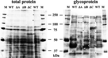
FIG. 7.
Comparison of soluble membrane proteins and glycoproteins. Proteins extracted with 1% Triton X-100 were loaded onto SDS-PAGE. Total proteins were stained with CBB, and the glycoproteins were detected by lectin blot analysis with concanavalin A as described in Materials and Methods. WT, ΔA, ΔB, and ΔC indicate proteins from wt, ΔAnpmtA, ΔAnpmtB, and ΔAnpmtC cells, respectively. Lanes M contained Precision plus protein standards used as molecular mass markers. Proteins that appeared in the wt but disappeared in the ΔAnpmt strain are indicated by black arrowheads; proteins that appeared in the ΔAnpmt strain but not in the wt are indicated by white arrowheads.

