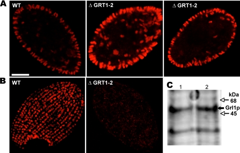
FIG. 5.
GRT1 and GRT2 are not essential for DCG formation and accumulation. (A) Visualization of docked DCGs by indirect immunofluorescence using a MAb (5E9) directed against the DCG lattice protein Grl3p. Shown are mid-cell sections of one wild-type (WT) and two ΔGRT1 ΔGRT2 (ΔGRT1-2) (cell line A) cells. Similar numbers of elongated DCGs are present, predominantly docked at the plasma membrane. (B) Visualization of docked DCGs by indirect immunofluorescence using a MAb (4D11) directed against Grt1p. Shown are tangential sections of one wild-type and one ΔGRT1 ΔGRT2 cell. The DCGs, seen en face, are docked at linearly arranged docking sites in the wild-type cell. No DCGs are seen in the ΔGRT1 ΔGRT2 cell since the MAb is directed against Grt1p. (C) Western blot analysis of SDS lysates of wild-type and ΔGRT1 ΔGRT2 (line A) cells using a polyclonal antibody against the granule lattice protein Grl1p. A total of 2 × 10⁵ cell equivalents were loaded for each sample, and the bound antibody was detected using the Odyssey system (see Materials and Methods). Lane 1, wild type; lane 2, ΔGRT1 ΔGRT2. Wild-type and ΔGRT1 ΔGRT2 cells accumulate similar levels of Grl1p.

