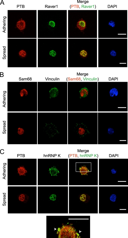
FIG. 3.
hnRNP K, but not Raver1 or Sam68, relocalizes to the cytoplasm during cell adhesion. MEFs were lifted and maintained in serum-free medium on a rotator for 1 h at 37°C and then replated on fibronectin-coated coverslips in the presence of serum. Cells were incubated for 15 min (adhering) or overnight (spread), fixed, and immunostained with antibodies to Raver1 and PTB (A), Sam68 and vinculin (B), or PTB and hnRNP K (C). Adhering cells were identified by cytoplasmic staining for PTB (A and C) or by punctate vinculin staining (B). The expanded view at the bottom shows a partial overlap between PTB and hnRNP K in the cytoplasm and periphery. Arrowheads indicate regions of colocalization. Scale bars, 20 μm.

