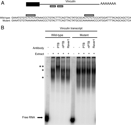
FIG. 7.
PTB binds CU repeats in the 3′ UTR of vinculin transcript. (A) Schematic showing potential PTB binding sites in the 3′ UTR of mouse vinculin mRNA. The black box indicates the carboxy-terminal coding region, the black line indicates the 3′ UTR, and the gray boxes highlight CU repeats. Transcripts synthesized for PTB binding experiments are shown below the schematic. Wild-type transcript corresponding to the CU repeat region (gray boxes) of the vinculin mRNA and a mutant transcript in which the CU repeats were changed to AA repeats are shown. (B) EMSA analysis of wild-type and mutant vinculin transcripts in HeLa nuclear extract. Labeled transcript was incubated with HeLa nuclear extract for 30 min and then incubated with the indicated antibodies to supershift RNA/protein complexes. Free RNA is shown at the bottom of the gel. A single asterisk identifies shifted RNP complexes, and the double asterisk identifies antibody-supershifted complexes.

