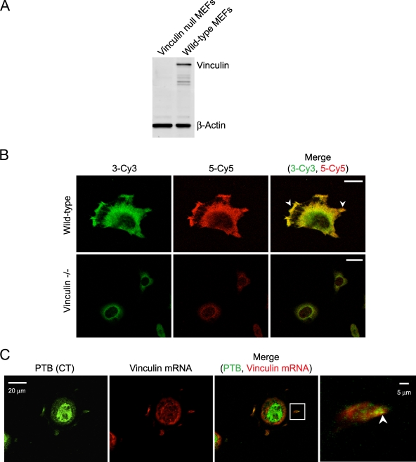
FIG. 8.
Vinculin mRNA colocalizes with PTB to the cell periphery of spreading fibroblasts. (A) Western blot for vinculin and beta-actin in wild-type and vinculin null MEFs shows an absence of vinculin protein in the null cells. (B) Wild-type and vinculin null MEFs were fixed with 4% PFA, and in situ hybridization was performed with two riboprobes specific to different regions of the 3′ UTR of vinculin mRNA. Riboprobe 3 was Cy3 labeled, and riboprobe 5 was Cy5 labeled. The arrowheads identify vinculin mRNA localization to protrusions and cell periphery. (C) Cells allowed to adhere for 20 min were fixed and immunofluorescence performed with anti-PTB (CT) antibody. Cells were postfixed with PFA, and in situ hybridization was performed with Cy3-labeled vinculin riboprobes 3 and 5 combined. The arrowhead in the enlarged image identifies regions of colocalization in newly formed protrusions. Scale bars, 20 μm except in the enlarged images to the right in panel C (5 μm).

