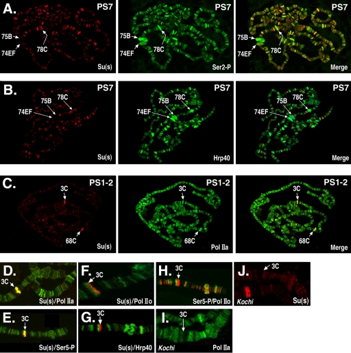
FIG. 1.
Su(s) localizes to a subset of Pol II-associated sites. The polytene chromosome distributions of Su(s) and either Pol II or Hrp40 were compared by immunofluorescence analysis. (A to C) Global Su(s) distribution at specific puff stages (PS). Arrows indicate the cytological positions of several prominent puffs. Chromosomes were probed with antibodies that recognize Su(s) (red) and either Pol II or Hrp40 (green). Sites of colocalization appear yellow in merged images. (D to J) Distribution of Su(s), various forms of Pol II, and Hrp40 at the 3C locus during PS 1 to 2. Genotypes: wild type (D to H); Kochi mutant with Sgs4 enhancer deletion (I and J). Panels D to H show merged images of chromosomes probed with the antibodies indicated in the lower right corner. The chromosomes shown in panels I and J were probed with a single antibody. Antibody signals are shown in the following colors: Su(s), red (D to H and J); Ser5∼P, green (E) and red (H); Pol IIa, green (D and I); Hrp40, green (G); and Pol IIo, green (F and H). Su(s) colocalizes at 3C with Pol IIa and Ser5∼P but not Ser2∼P or Hrp40. Pol IIa and Su(s) are absent from 3C in the Kochi mutant.

