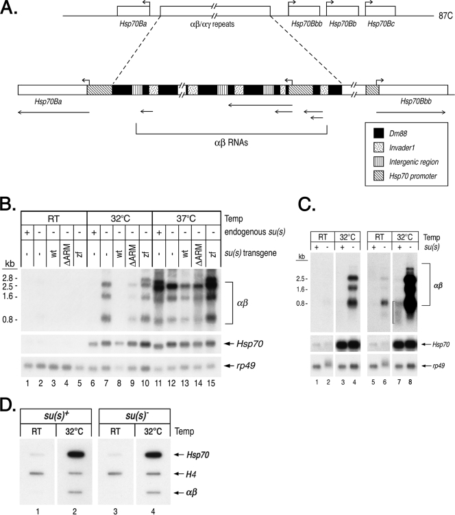
FIG. 4.
Su(s) inhibits the accumulation of noncoding αβ RNAs from the 87C region during a mild heat shock. (A) Schematic map of transcription units in the 87C region, which contains several Hsp70 genes and a cluster of tandemly repeated αβ/αγ elements. The αβ elements are segments of transposons invader1 and Dm88, whereas a γ element is a duplicated Hsp70 promoter fragment joined to αβ sequences. Hsp70 transcription start sites are indicated by the right-angled arrows above the map. Regions that give rise to αβ and Hsp70 RNAs are indicated by lines with arrows beneath the map. (B, C) Northern blots of RNA from heat-shocked and control (RT) flies. The abundance of αβ RNA is substantially lower than Hsp70 RNA; thus, blots probed to detect αβ RNAs were exposed for longer times. (B) RNA from su(s)+, the su(s) mutant, and flies carrying the su(s) transgenes indicated in the null mutant background. (C) RNA from su(s)+ and the su(s) null mutant. Two different exposures of the blot probed for αβ RNA are shown in lanes 1 to 4 and lanes 5 to 8. The bracket in lane 7 indicates the residual, heterogenous-sized αβ transcripts seen in su(s)+ RNA at 32°C. (D) Nuclear run-on experiment comparing the amount of elongating Pol II in the αβ/αγ region after a 32°C heat shock in wild-type and su(s) mutant adult flies. Internal control histone H4 genes are transcribed in the presence and absence of heat shock. Probes in panels B to D are indicated on the right. This analysis indicates that Su(s) does not regulate the transcription of the αβ/αγ region. wt, wild type.

