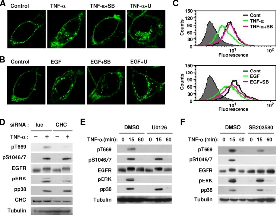
FIG. 5.
Phosphorylation-dependent endocytosis and subsequent dephosphorylation of EGFR. (A and B) HeLa-EGFR-GFP cells were pretreated with SB203580 (SB) (10 μM) and U0126 (U) (5 μM) for 30 min and then stimulated with TNF-α (A) or EGF (B) for another 15 min. Subcellular localization of EGFR-GFP was examined by confocal fluorescent microscopy. (C) Cells were pretreated with SB203580 (10 μM) for 30 min and then stimulated with 20 ng/ml TNF-α or 10 ng/ml EGF for 15 min. Cell surface expression of EGFR was investigated by FACS analysis. Cont, control. (D) HeLa cells were transfected with siRNAs against CHC and Luc. At 72 h posttransfection, cells were stimulated with TNF-α for 10 min. Whole-cell lysates were immunoblotted with the indicated antibodies. (E and F) HeLa cells were pretreated with U0126 (5 μM) (E) or SB203580 (10 μM) (F) for 30 min and then stimulated with TNF-α for another 15 and 60 min. Whole-cell lysates were immunoblotted with the indicated antibodies. DMSO, dimethyl sulfoxide.

