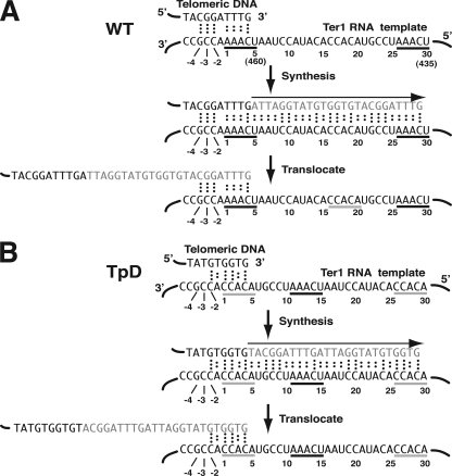
FIG. 1.
Putative base-pairing interactions between the telomere and the telomerase RNA template region of K. lactis wild type (WT) and the TpD template permutation mutant. (A) Diagram of telomeric repeat synthesis using a wild-type TER1 template. The top panel shows potential base-pairing interactions between a single-stranded end of telomeric DNA with the region immediately 3′ to the template of the wild-type Ter1 RNA. Dots between bases indicate hydrogen bonds of base pairs. Numbers indicate nomenclature used for base positions in this work. Numbers in parentheses indicate base positions numbered from the 5′ end of the Ter1 RNA. The middle panel shows the synthesis step when a 25-nt telomeric repeat (shown in gray) is copied. The bottom panel shows the same sequences after translocation of the newly synthesized DNA such that the new 3′ DNA end is base paired back at the 3′ end of the template. Sequences underlined in black indicate the 5-nt direct repeats of the template region. The sequence underlined in gray indicates the 5-nt sequence engineered to be the direct repeats of the template region in the TER1-TpD permutation mutant. (B) Diagram of telomeric repeat synthesis using a TER1-TpD template. The TER1-TpD mutant has a 30-nt template region that is circularly permuted relative to the normal template. The sequences underlined in gray are the novel 5-nt repeats, and the sequence underlined in black shows the position of the sequence that comprises the 5-nt repeats (bases 1 to 5 and 26 to 30) in the wild-type template. Aside from different start and stop points for DNA synthesis, the TpD template is expected to produce wild-type telomeric repeats. The region from −1 to −4 next to the template is identical in wild-type Ter1 and Ter1-TpD, but as shown, that region is expected to interact with different parts of the telomeric repeat in the two cell types.

