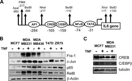
FIG. 2.
Weakly and highly invasive breast cancer cell lines demonstrate overexpression of NF-κB and AP-1 transcription factor families. (A) Schematic representation of the highly conserved IL-6 promoter region with indication of the various embedded transcription factor and restriction enzyme motifs. (B) On total cell lysates of MCF7, MDA-MB231, MDA-MB468, T47D, and ZR75 cells, left untreated or treated for 30 min with TNF, we performed Western analysis against Fra-1, c-Jun, NF-κB p65, RelB, and β-actin, as indicated. (C) On total cell lysates of MCF7 or MDA-MB231 cells, left untreated or treated for 30 min with TNF, we performed Western analysis against CREB, C/EBPβ, and tubulin.

