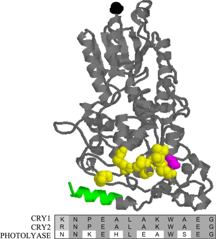
FIG. 8.
CRY2G354 is immediately upstream of a region of amino acids conserved in repressive CRYs but not Photolyase. CRY2 3D homology model. This model was generated as described for Fig. 2. The N-terminal residue is shown in black, while the coiled-coil domain is shown in green. CRY2G351 is drawn in magenta, while the residues in the region adjacent to G351 that are conserved in all repressive CRYs, but not Photolyase, are shown in yellow. Under the model is a protein alignment of these residues in CRY1, CRY2, and Photolyase. Conserved residues are shaded in gray.

