FIG. 3.
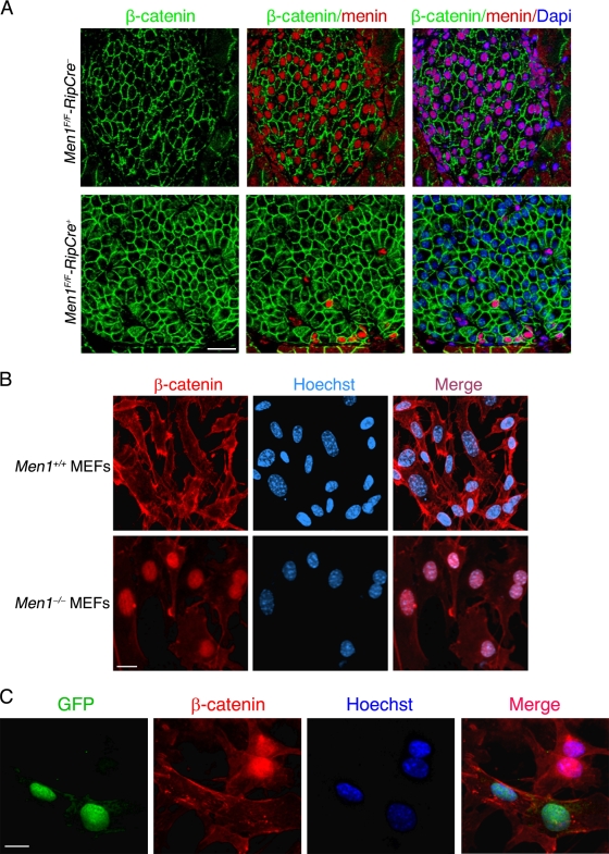
Nuclear accumulation of β-catenin in the absence of menin. (A) Pancreatic islets obtained from 6-month-old Men1F/F-RipCre− and Men1F/F-RipCre+ mice. Pancreas sections were stained with β-catenin antibody (green) and menin antibody (red). Nuclei were stained with DAPI (4′,6′-diamidino-2-phenylindole) (blue). (B) In menin-null MEFs (Men1−/−), β-catenin (red) is largely accumulated in the nucleus, in contrast with cytoplasmic membrane localization in wild-type (Men1+/+) MEFs. Nuclei are stained with Hoechst stain (blue). (C) Translocation of nuclear β-catenin. Men1−/− MEFs were transfected with menin-GFP. Cells were stained with β-catenin antibody (red). Nuclei are stained with Hoechst stain (blue). Scale bars, 30 μm.

