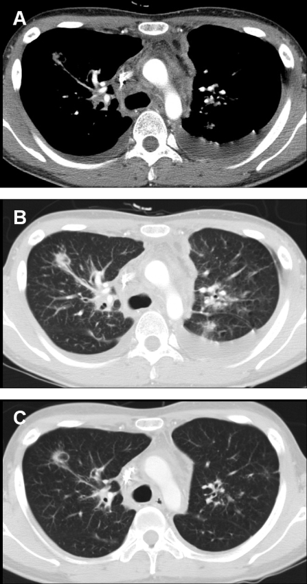
FIG. 1.
Twenty-three-year-old man with disseminated Mycobacterium bolletii infection. (A) A contrast-enhanced chest CT scan obtained at the level of the carina by using the mediastinal window setting shows extensive mediastinal lymphadenopathy. Note the bilateral pleural effusion. (B) A chest CT scan obtained using the lung window setting shows multiple pulmonary nodules. (C) After 4 weeks of antibiotic therapy, the chest lesions, including the mediastinal lymphadenopathy and parenchymal nodules, were improved. Also note the disappearance of the bilateral pleural effusion.

