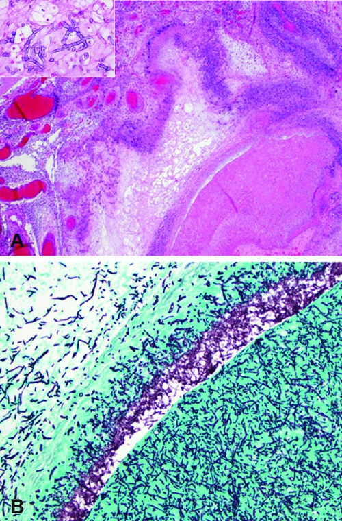
FIG. 1.
(A) The thrombosed left middle cerebral artery was surrounded by granulomatous inflammation in the meninges (hematoxylin and eosin; original magnification, ×20). (Inset) The fungal hyphae were nonpigmented and septate (hematoxylin and eosin; original magnification, ×400). (B) Silver stain highlights the fungal hyphae within the thrombus and extending through the arterial wall (Gomori methenamine silver; 100× original magnification).

