Fig. 2.
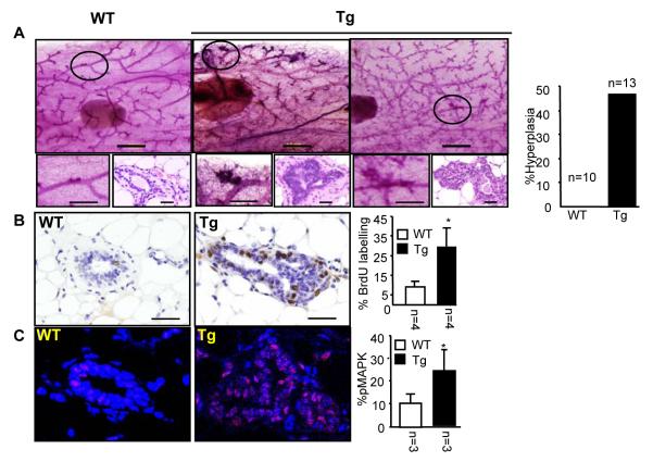
Cytoplasmic PELP1 in mammary epithelium causes hyperplasia and promotes non-genomic signaling. (A) Whole-mount and histological analyses of MMTV—cyto-PELP1 transgenic mice (Tg) showing the mammary hyperplasia at 12 weeks of age compared with age-matched wild-type (WT) mice. (B) BrdU-positive cells for both transgenic and WT mice counted and expressed as a ratio of positive nuclei to the total number of nuclei counted (percentage). (C) Immunofluorescence analysis with anti—phospho-MAPK antibody showing increased activation of the MAPK (red) in transgenic glands (TG) compared with WT control glands. Nuclei were stained with DAPI (blue). The plot on the right panel shows the quantitative analysis of percentage of positive cells for phospho-MAPK.

