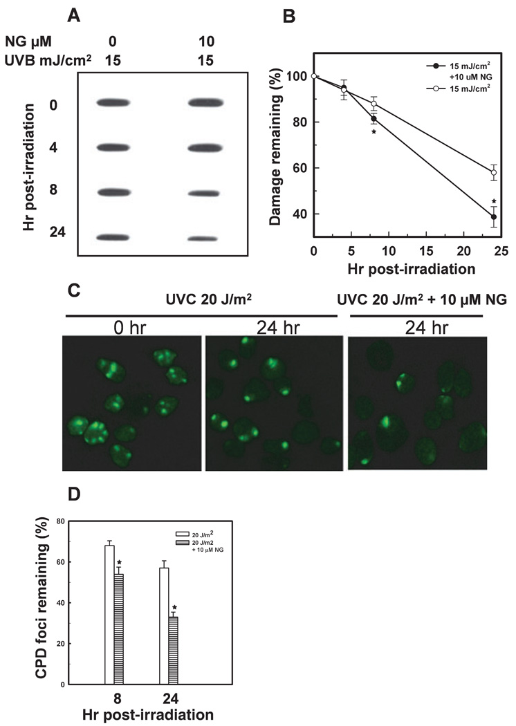
Figure 7.
NG enhances the removal of CPD from the genome of HaCaT cells. Cells were grown to 100% confluency in plates or on coverslips, left unirradiated or irradiated with UVB or UVC, and incubated in fresh medium with or without NG. The cells were allowed to repair the damage for the indicated times. The CPD in genomic DNA were quantitated by immunoslot-blot assay using CPD damage-specific antibodies. A representative plot and mean ± SD of five independent experiments are shown (A and B). For immunofluorescence detection of CPD foci, cells were irradiated with 20 J m−2 UVC through an isopore filter. The cells were allowed to repair the damage, fixed with 2% paraformaldehyde and immunostained with anti-CPD antibody (C and D). CPD foci in 200 individual cells from three different microscope fields were counted for calculating the percentage of remaining damage. *Shows significant difference from UV-treated cells at P < 0.05.

