
Figure 1.
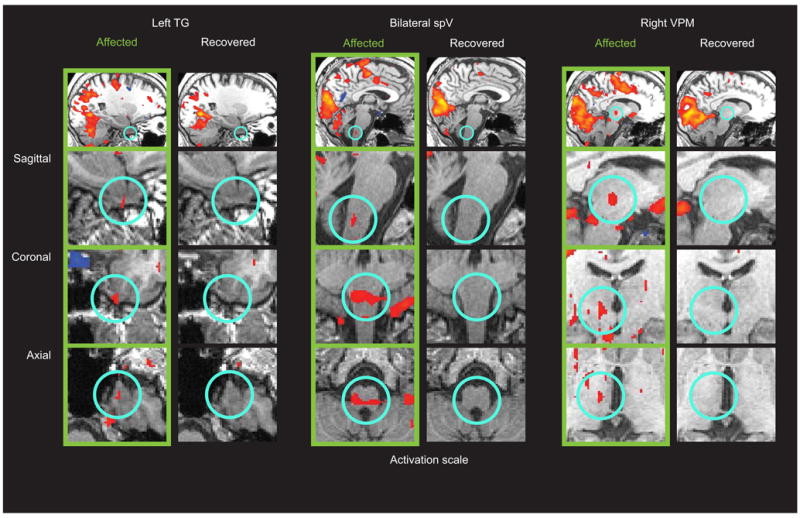
fMRI activation during photophobia across three levels of the trigeminal system. Scanning results from during the photophobic state (“Affected” in green) and after recovery (“Recovered”) are shown. Regions of interest (highlighted by light blue circles) are shown across three different viewing planes. Significant activation (p<0.0001, uncorrected) was detected in the “Affected” state in left trigeminal ganglion (TG), bilateral trigeminal nucleus caudalis (spV), and right ventroposteromedial thalamus (VPM). No significant activation in these regions was detected in the “Recovered” state. Other activations of note include the occipital gyrus and anterior cingulate cortex.