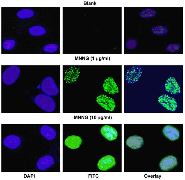
Fig. 2. MNNG induces γH2AX foci formation in FL cells.
After MNNG treatment for 8 h, cells were fixed and stained with anti-γH2AX antibody, and subjected to immunofluorescent microscopy. Shown are representative images from one of four independent experiments. Upper panel, control; middle and lower panel, MNNG treated cells. Blue, DAPI stain for nuclei; green, γH2AX. Notice that γH2AX all present in the nuclei. (For interpretation of the references in color in this figure legend, the reader is referred to the web version of this article.)

