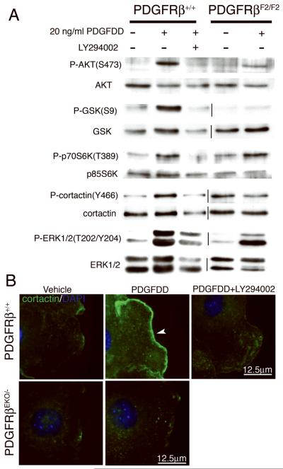
Figure 7. Disruption of PDGF signaling results in failure to activate and localize cortactin.
(A) Western blot analysis of primary epicardial cell lysates from the indicated genotypes and treatments were probed for phosphorylated (top panel) and total protein (bottom panel). (B) Cortactin localization on wounded epicardial cells. Cells were stimulated with 50ng/ml PDGFDD. Arrowhead indicates abundant cortactin localization to lamellipodia.

