
Figure 4.
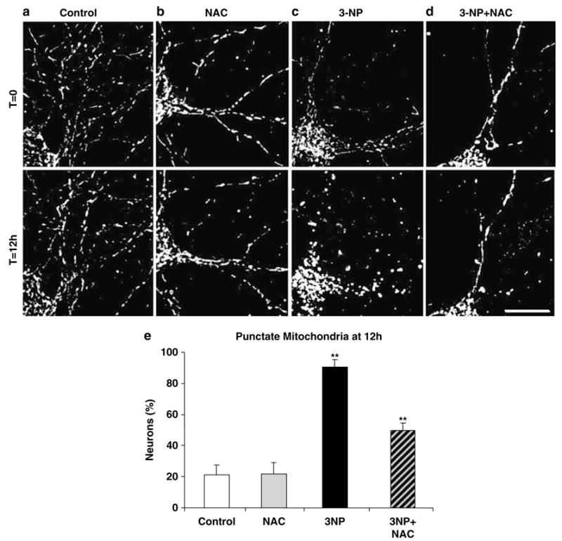
Oxidative and nitrosative stress inhibition alleviates 3-NP-induced formation of punctate mitochondria. Representative snap shots of a time-lapse series of a DsRed2-Mito-transfected control neuron (a), a 3-NP (10 mM) exposed neuron (b), a neuron pretreated 3 h with NAC (50 μM) followed by 3-NP (10 mM) exposure (c), and a neuron pretreated with NAC alone (50 μM) (d) – all at time zero (T =0, upper) and 12 h later (T = 12 h, lower). Scale bar, 10 μm. (e) Bar graph of the percentage of neurons with punctate mitochondria at 12 h of 3-NP exposure. Data represent four independent experiments and error bars indicate ± S.E.M. Significant differences are indicated by symbols **(P<0.01; one-way ANOVA followed by Fisher’s t-test)