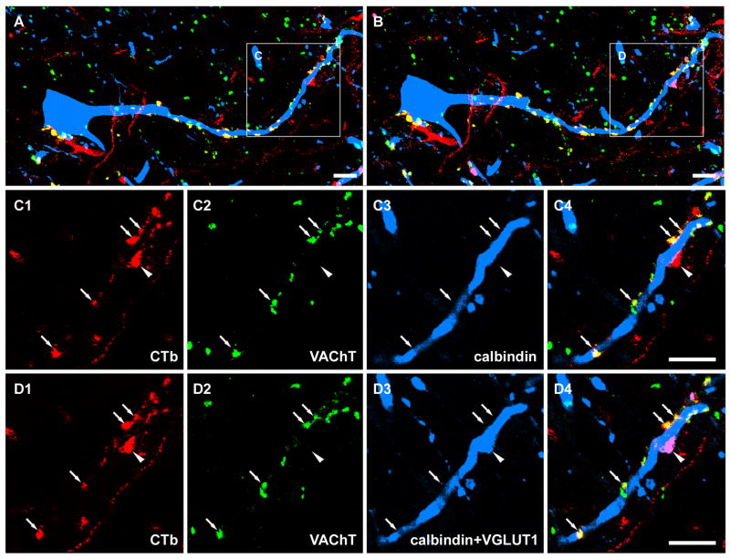
Figure 4.
Sequential immunocytochemistry for CTb labelled terminals in contact with a Renshaw cell. A and B show a general overview of CTb labelled terminals in contact with a calbindin labelled cell before and after a reaction with a fourth antibody against VGLUT1. Details of the areas demarcated by the boxes are shown in C1-C4 and D1-D4. C1-C4 are single optical sections illustrating that most of the CTb labelled terminals in contact with the calbindin cell were positive for VAChT. Arrows indicate double labelled CTb axon terminals with VAChT staining. The arrowhead indicates a single CTb labelled terminal on the Renshaw cell that was negative for VAChT. D1-D4 are single optical sections of the same terminals that were rescanned after sequential incubation with a fourth antibody against VGLUT1. The extra labelling present in D3 (indicated by arrowhead), which was absent in C3, corresponds to additional VGLUT1-immunostaining (see D4). Note that this VGLUT1 positive terminal, which forms an apposition with the Renshaw cell, is bigger than CTb labelled motoneuron terminals, and is likely to be a primary afferent terminal. A, B, C4 and D4 are merged images. Scale bar: 1μm.

