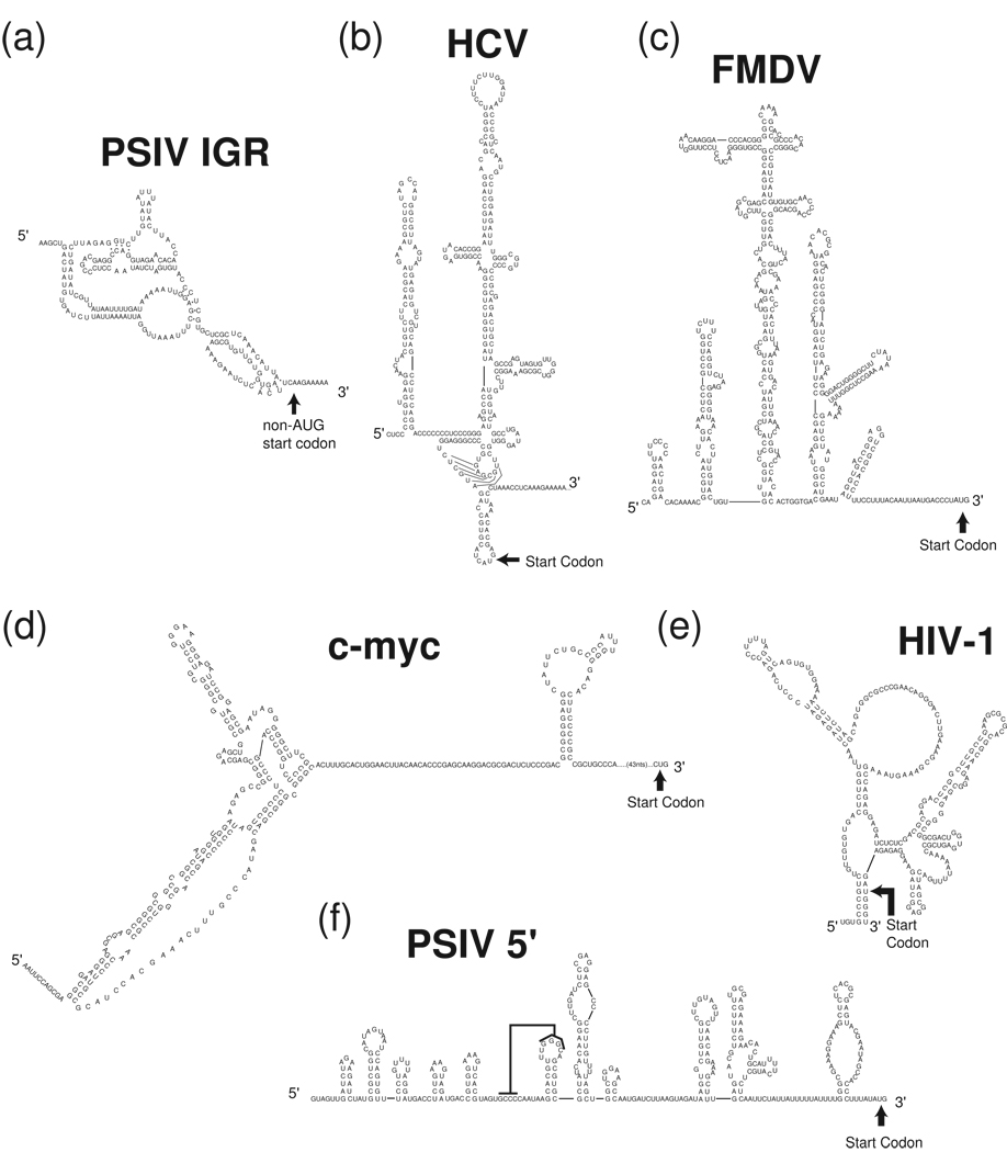
Figure 3. Examples of viral and cellular IRES secondary structures.
Experimentally tested secondary structures of several diverse viral and cellular IRES RNAs are shown. (a) Plautia stali intestine virus (PSIV) IGR IRES. (b) HCV IRES (c) FMDV IRES (d) c-myc IRES (e) Human immunodeficiency virus-1 (HIV-1) gag- IRES (f) PSIV 5' IRES, the black line indicates a proposed pseudoknot interaction. Note that these secondary structures may be revised as more information becomes available regarding differences between RNA made and folded in vitro versus that made in vivo, which folds co-transcriptionally.

