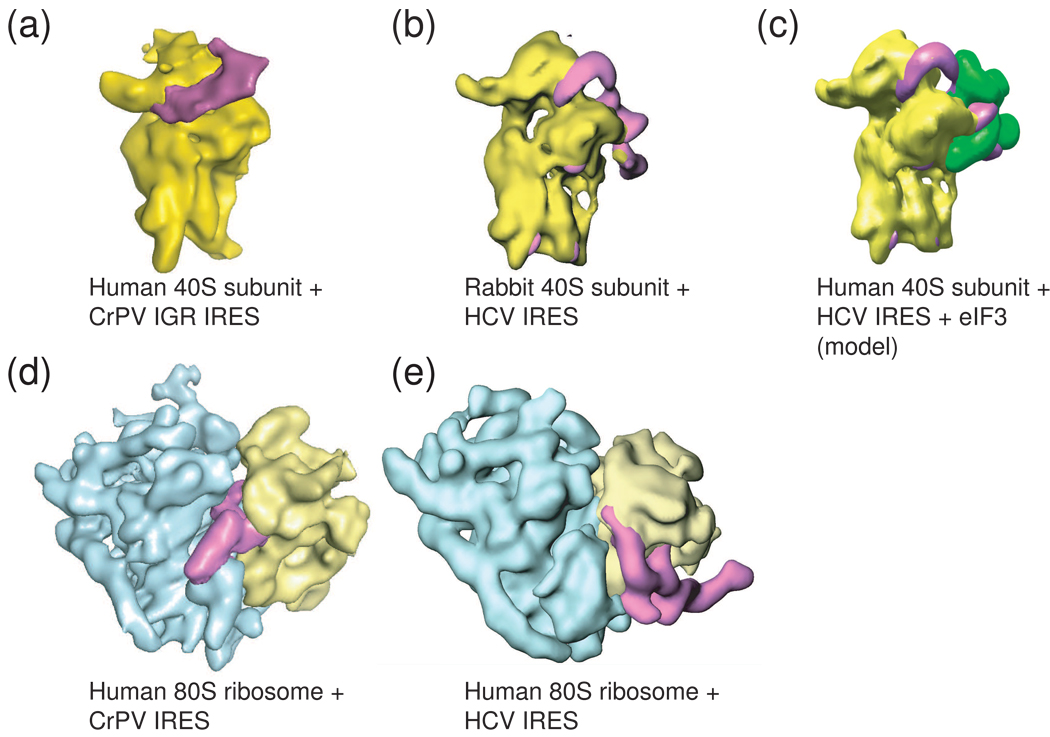
Figure 4. Cryo-EM reconstructions of two different viral IRESs bound to the ribosome.
To date, only the HCV IRES (and related) and the Dicistroviridae IRES RNAs have been shown to bind directly to ribosomal subunits, and these complexes have been studies by cryo-EM. (a) The Cricket paralysis virus (CrPV) IGR IRES (magenta) bound to the 40S subunit (yellow). (b) The HCV IRES (magenta) bound to a 40S subunit (yellow). These two IRES types occupy different sites on the subunit, but both induce a similar conformational change in the 40S subunit when they bind, suggesting some mechanistic convergence. (c) Model of the HCV IRES bound to the 40S subunit and eIF3 (green), built from several cryo-EM reconstructions. (d) The CrPV IGR IRES bound to an 80S ribosome, the view is rotated 90 degrees from the view in panel (a). Note that higher resolution reconstructions of this complex have been published, but to allow more direct comparison with the HCV IRES-bound ribosome, the middle resolution structure is shown. (e) The HCV IRES bound to an 80S ribosome. Again, the different binding modes of these two IRESs to the ribosome are obvious, as are differences in the overall folded architectures of the IRESs; the CrPV IGR IRES is more compact, while the HCV IRES is extended. Cryo-EM reconstructions of preiniation complexes containing other IRESes have not been reported.

