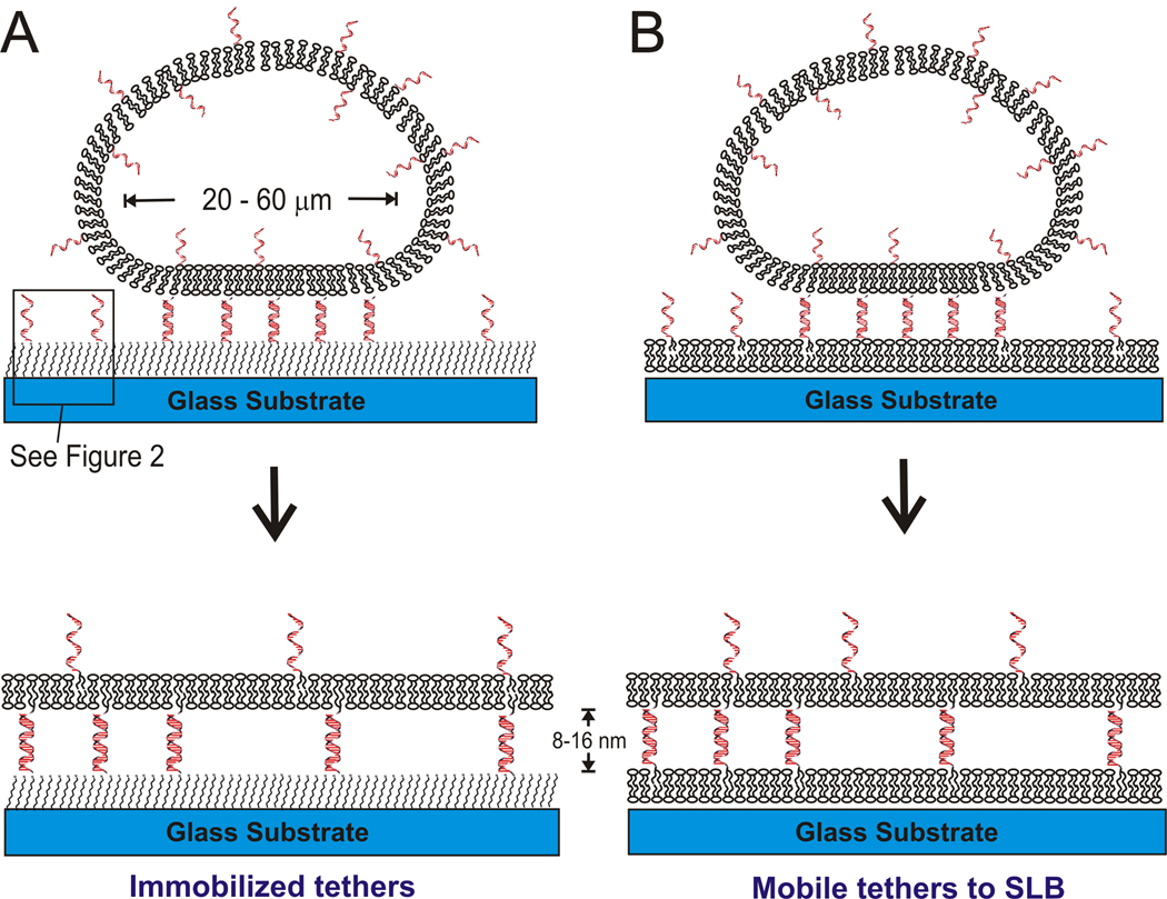
Figure 1.
schematic diagram of DNA-tethered membrane formation by GUV rupture onto (A) self-assembled alkylsilane monolayers, see Figure 2 for details; and (B) supported lipid bilayers. The GUV membrane contains DNA-linked lipids, which mediate vesicle binding and rupture. In (A), the glass substrate is functionalized with an azide-terminated alkyl-siloxane monolayer to which complementary DNAs are immobilized via the click reaction (see Figure 2). The distance from the substrate is controllable by adjusting the DNA length, about 8 nm for 24mer and 16 nm for 48mer DNA. In (B), a supported lipid bilayer presenting DNA is first formed on the glass substrate, and then exposed to GUVs presenting the complementary DNA to form a DNA-tethered membrane. In both cases, tethered membrane patches whose area is approximately that of the original GUVs are formed.

