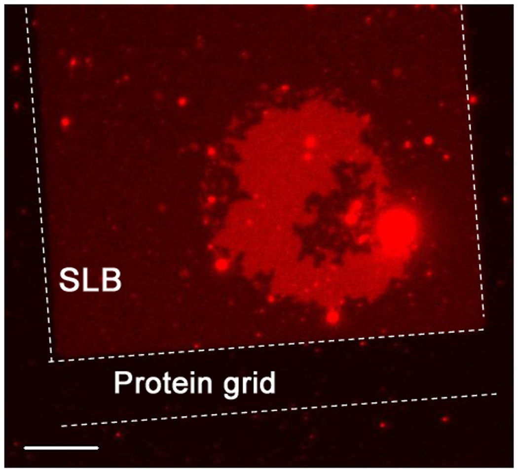
figure 5.

A fluorescently labeled tethered bilayer patch (bright red) with mobile DNA tethers on an unlabeled supported bilayer in the process of disintegrating. The supported bilayers are confined by a protein grid outlined with dotted lines, to observe the build-up of weak red fluorescence from the lipid materials that were part of the unstable tethered patch. This shows that the broken part of the tethered bilayer remains mostly on the SLB. One side of the rectangular protein grid is 100 µm and the scale bar is 15 µm.
