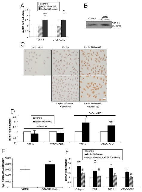
Figure 4. Gene and protein expression of TGFβ1 and CTGF/CCN2 mediated by leptin in cultured KCs.
All KCs were cultured for 48 h prior to treatment. Three independent experiments were performed. (A) mRNA expression of TGFβ1 and CTGF/CCN2 in wt rat KCs cultured for 24 h in the presence of leptin (10 or 100 nmol/L). (B) Immunoblot of TGFβ1 in KC-conditioned medium treated with leptin 100 nmol/L for 24 h. (C) Immunocytochemical determination of CTGF/CCN2 protein in KCs. KCs were cultured for 24 h with leptin 100 nmol/L, leptin 100 nmol/L + sTGFβR fusion protein (50 μg/mL) or leptin 100 nmol/L + human IgG (50 μg/mL). Scale bar: 20 μm. (D) mRNA expression of TGFβ1 and CTGF/CCN2 in Zucker (fa/fa) and lean (Fa/Fa) rat KCs cultured for 24 h in the presence of leptin (100 nmol/L). (E). KC intracellular H2O2 generation by leptin. Leptin 100 nmol/L was incubated with KC (48 h cultured) for 24 h. The excitation wavelength was 485 nm and emission wavelength 535nm. (F). Collagen I, TIMP1, TGFβ1 and CTGF/CCN2 mRNA expression in HSCs incubated with KC-conditioned medium in the presence or absence of TGFβ antibody (10 μg/mL).* P<0.05 and ** P<0.01 compared to control group. # p<0.05 compared to the leptin treatment group without TGFβ antibody.

