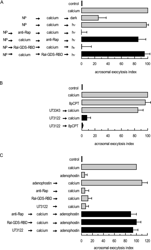
FIGURE 3.
Rap1 participates in a pathway that mobilizes calcium from an IP3-sensitive store. A, permeabilized spermatozoa were loaded with 10 μm NP-EGTA-AM (NP) for 10 min at 37 °C to chelate intra-acrosomal calcium. The AR was subsequently initiated by adding 0.5 mm CaCl2. After a further 15-min incubation at 37 °C to allow exocytosis to proceed up to the intra-acrosomal calcium-sensitive step, sperm were treated for 15 min at 37 °C with 20 μg/ml anti-Rap antibody (Santa Cruz Biotechnology) or 1 μg/ml Ral-GDS-RBD. All these procedures were carried out in the dark. UV flash photolysis of the chelator was induced at the end of the incubation period (hv), and the samples were incubated for 5 min (NP → calcium → inhibitor → hv; black bars). Several controls were run (gray bars) as follows: background AR in the absence of any stimulation (control); AR stimulated by 0.5 mm CaCl2 (calcium), inhibitory effect of NP-EGTA-AM in the dark (NP → calcium → dark), and the recovery upon illumination (NP → calcium → hv); and the effect of the inhibitors when present throughout the experiment (NP → inhibitor → calcium → hv). Sperm were fixed; the AR was measured by FITC-PSA binding, and the data were normalized as described under “Experimental Procedures.” B, permeabilized spermatozoa were incubated with 15 μm U73343 or U73122 for 15 min at 37 °C. Acrosomal exocytosis was then initiated by adding 0.5 mm CaCl2 (calcium) or 50 μm 8-pCPT-2′-O-Me-cAMP (8pCPT) and a further 15-min incubation at 37 °C (black bars). Sperm were stained, and the AR scored as in A. C, permeabilized spermatozoa were incubated with 20 μg/ml anti-Rap antibodies (Santa Cruz Biotechnology), 1 μg/ml Ral-GDS-RBD, or 15 μm U73122 for 10 min at 37 °C. Exocytosis was initiated by adding 0.5 mm CaCl2. To demonstrate that Rap1-GTP and an active PLC were located upstream of the intracellular calcium efflux, 5 μm adenophostin was added to mobilize calcium from IP3-sensitive stores, and incubations proceeded for an additional 10 min at 37 °C (black bars). Controls (gray bars) included the following: background AR in the absence of any stimulation (control), AR stimulated by 0.5 mm CaCl2 (calcium), AR inhibition by 20 μg/ml anti-Rap antibodies, 1 μg/ml Ral-GDS-RBD, or 15 μm U73122, and AR unaffected by 5 μm adenophostin. Sperm were stained, and the AR scored as in A. Plotted results represent the mean ± S.E. of at least three independent experiments.

