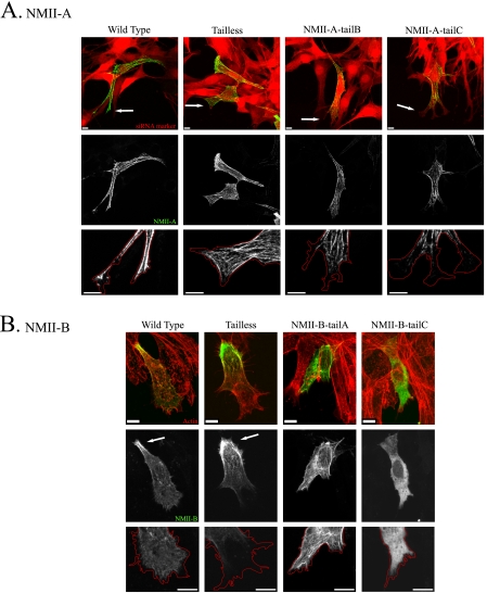
FIGURE 4.
NMII-A and NMII-B tailpiece effect on isoform localization in vivo. Tailpiece-swapped full-length NMII-A and NMII-B fused to GFP were transfected into cells and subjected to wound scratch assay as described under “Experimental Procedures.” A, NMII-A tail-swapped chimeras expressed in NMII-A siRNA-treated B−/B− MEF cells. Arrows point at cells leading edge. Red, NMII-A siRNA marker; green, GFP-NMII-A. B, NMII-B tail-swapped chimeras expressed in B−/B− MEF cells, actin was detected by Rhodamine-Phalloidin staining. Arrows indicate posterior accumulation of NMII-B. Red, actin; green, NMII-B. Red lines are tracing of cells leading edge. Bar = 10 μm.

