FIGURE 4.
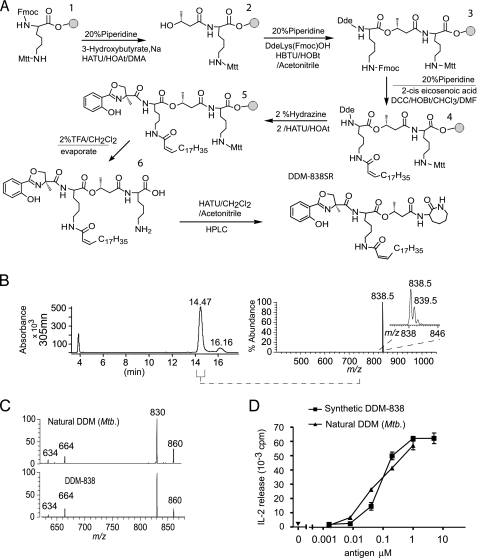
Synthetic DDM-838SR is indistinguishable from M. tuberculosis DDM-838. A, a schematic of solid phase synthesis of DDM-838 is shown with the gray circle representing super acid-sensitive resin. B, HPLC with UV and MS detection was used to collect the fraction from 14.1 to 14.7 min using a split interface for diversion to the mass spectrometer. Positive mode electrospray ionization-MS showed the expected mass and isotope pattern for [M + H]+ of DDM and no evidence of significant contamination by other products. C, MS-MS analysis of M. tuberculosis and synthetic DDM-838SR [M + Na+] ion showed spectra that were nearly identical. The fragment at m/z 830 resulting from cleavage through the oxazoline ring (see supplemental Fig. S1) is clearly observed with similar signal intensity in the spectra of both compounds, indicating that this functional group was unaltered under the conditions used for synthesis and isolation. D, CD1a-restricted T cell activation was measured by interleukin-2 release in response to human monocyte-derived dendritic cells and DDM from synthetic and natural sources.

