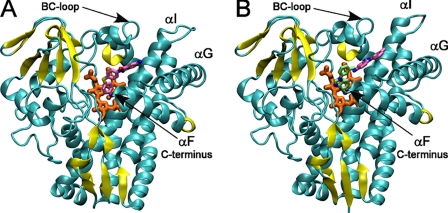
FIGURE 3.
Overall view of CYP130 with ligands bound in the active site. A, compound 2 is highlighted in pink. B, two molecules of compound 4 are highlighted in lime and pink. Protein is shown as a ribbon with the β-sheets highlighted in yellow, and heme is in orange. Images were generated using the Visual Molecular Dynamics (VMD) program (28).

