
Figure 2.
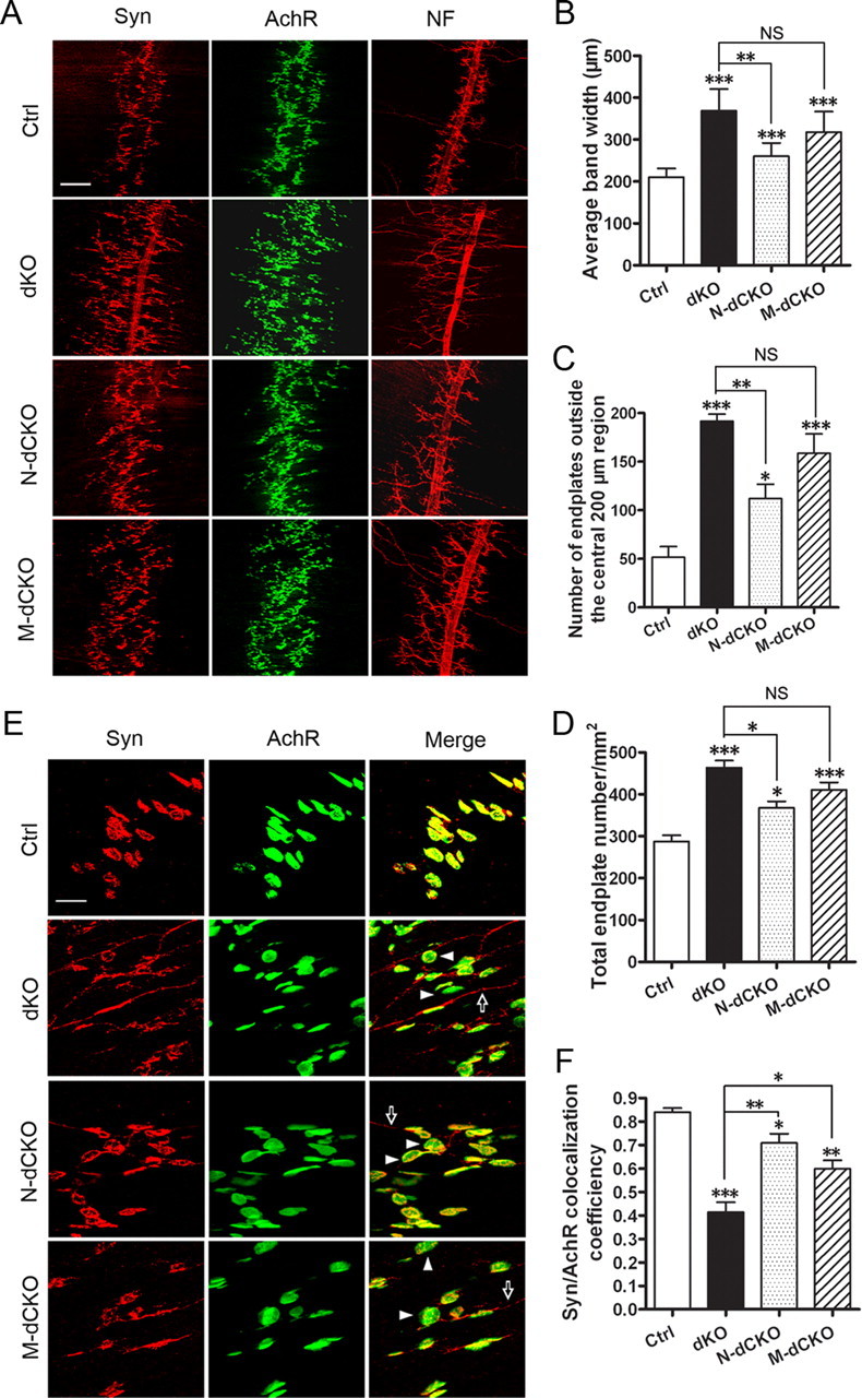
Neuromuscular synapse defects in APP/APLP2 motor neuron (N-dCKO) and muscle (M-dCKO) knock-out mice. A, Whole-mount immunostaining of control, dKO, N-dCKO, or M-dCKO P0 diaphragm muscles with antibodies against Syn or NF. α-BTX staining was used to mark the AchRs. Both the germline and conditional mutants showed diffused presynaptic and postsynaptic distribution and nerve terminal sprouting. B, Quantification of the average band width of AchR-positive endplates. C, Quantification of the number of AchR-positive endplates outside the central 200 μm band zone. D, Measurement of the number of endplate per unit area. B–D are expressed as mean ± SEM (6 animals/genotype). E, Higher magnification images of synapse structures with representative endplates poorly covered by Syn marked by arrowheads and extrasynaptic Syn staining by arrows. F, Quantification of the percentage of AchR-positive endplates covered by Syn (average ± SEM of 20 endplates/genotype). The asterisks above each bar are in comparison with the control. The asterisks on top of the brackets are in comparison with the dKO mice. *p < 0.05; **p < 0.01; ***p < 0.001; NS, nonsignificant (p > 0.05) (one-way ANOVA). Scale bars: A, 100 μm; E, 20 μm.