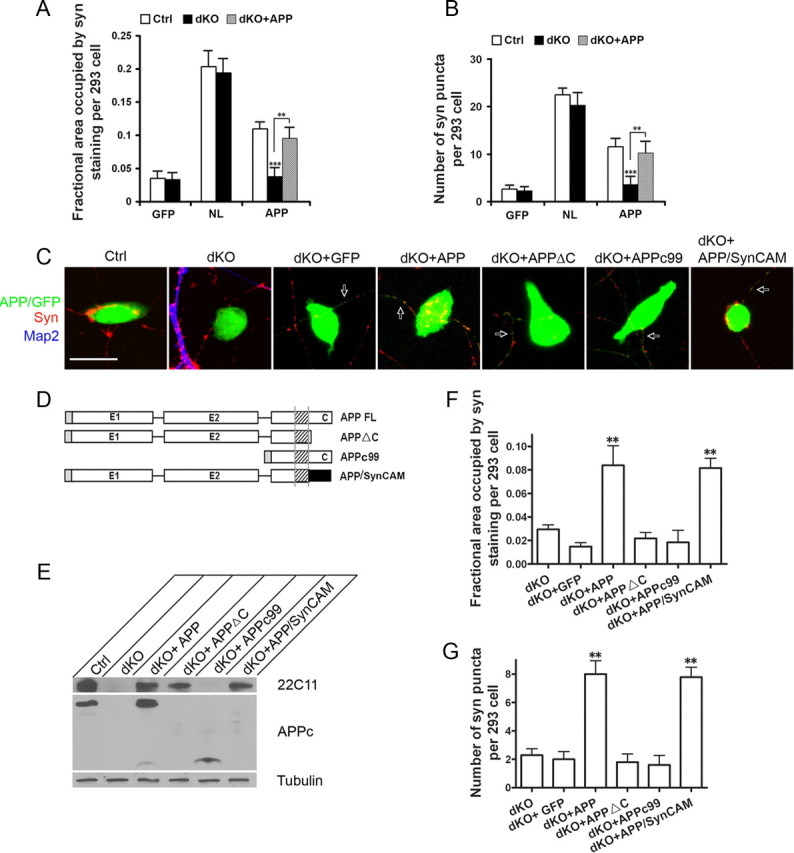
Figure 7.

Effect of neuronal APP on APP-induced synaptic puncta. The APP/APLP2-null neurons (dKO) and littermate APLP2-null controls (Ctrl) were cocultured with GFP, NL, or APP transfected HEK293 cells. dKO + APP, dKO neurons infected with APP-expressing lentivirus. The average area of transfect HEK293 cells covered by synaptophysin (A) and the average number of synaptic puncta per HEK293 cell (B) were quantified. APP-induced puncta is significantly reduced when cocultured with dKO neurons (Ctrl vs dKO, p < 0.001) (t test). This impairment is completely rescued by neuronal expression of APP (Ctrl vs dKO + APP, nonsignificant, p > 0.05). Error bars indicate SEM. C, Representative images of control (Ctrl), APP/APLP2 double knock-out (dKO), or dKO with lentiviral expression of GFP vector (dKO + GFP), human full-length APP (dKO + APP), intracellular domain deleted APP (dKO + APPΔC), extracellular sequence deleted APP (dKO + APPc99), or the APP/SynCAM chimera (dKO + SynCAM) cocultured with APP-transfected HEK293 cells. The cultures were stained with anti-synaptophysin (Syn; red) and anti-MAP2 (Map2; blue) antibodies. Both the transfected HEK293 cells and APP infected axons (arrows) are GFP-positive. Scale bar, 20 μm. D, Schematic diagram of the APP constructs. Black rectangle, SynCAM C-terminal sequences (amino acids 428-474). E, Western blot analysis of neuronal lysates from APLP2−/− control, dKO, and dKO neurons infected with APP and derivatives using the N-terminal and C-terminal antibodies 22C11 and APPc, respectively. Tubulin was used as a loading control. F, Quantification of the average area of HEK293 cells covered by synaptophysin immunoreactivity. G, Quantification of average number of Syn-positive puncta per transfected HEK293 cell. **p < 0.01 (t test) in comparison with the dKO + GFP control.
