
Figure 3.
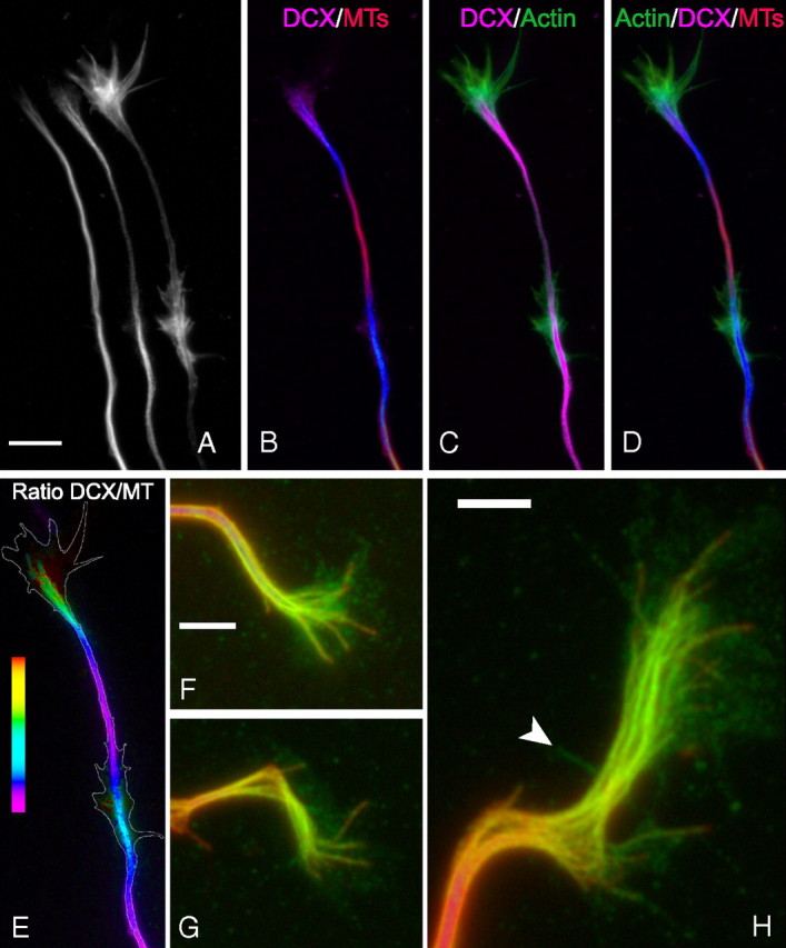
Higher-resolution analyses of microtubules, DCX, and actin staining in growth cones and focal axonal accumulations of DCX. The figure shows the distal axon and growth cone of cultured hippocampal neurons. A shows, from left to right, images of microtubule (MT), DCX, and actin filament staining. B–D show overlay images as indicated, with microtubule staining in red, DCX staining in violet, and actin staining in green. DCX staining is strong in the growth cone and at a site along the axon a bit proximal to the growth cone (these accumulations appear blue in the DCX/microtubule overlay image). Within the growth cone, DCX staining primarily colocalizes with microtubules, and the polymer rich in DCX extends into the actin-rich region of the growth cone. Note also that the region of the axon with the focal accumulation of DCX is also rich in actin filaments that form prominent lamellipodia. E shows a ratio image depicting the abundance of DCX relative to microtubules; the white outline represents the contour of actin staining. In the growth cone, the ratio increases progressively from the axon–growth cone neck region to the distal extent of the microtubule array (for a ratio image of another neuron, see Fig. 6). The ratio of DCX staining to microtubule staining is also elevated within the focal accumulation of DCX along the axon. Scale bar (in A): A–E, 7.2 μm. F–G show zoomed images of DCX (in green) and microtubule (in red) staining in three different growth cones. The colocalization of DCX with microtubules is readily apparent. There is also glow in the DCX channel that is not microtubule associated. Note that nonlinear enhancing methods were used to accentuate this glow without saturating that associated with microtubules. See Results for discussion of the possible significance of this non-microtubule fluorescence. The arrowhead in H identifies a filopodia detected by phalloidin staining (data not shown). Scale bars, 3.2 μm.