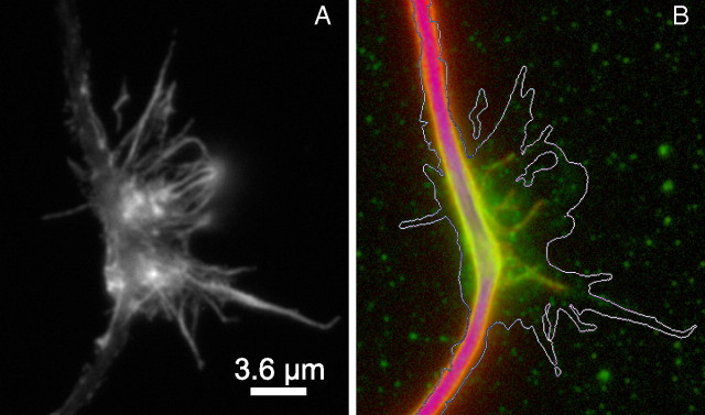
Figure 5.
Splayed microtubules at sites of focal accumulations of DCX along the axon. A shows actin filament staining associated with a focal accumulation of DCX. B shows an overlay of DCX (green) and microtubule (red) staining in the same region; the white line is an approximate outline of the actin staining. Note that several microtubules splay out from the main microtubule bundle and that these are decorated with DCX. Nonlinear methods were used to enhance the staining associated with the splayed microtubules without grossly saturating the staining associate with the microtubule bundle of the axonal shaft. Scale bar, 3.6 μm.

