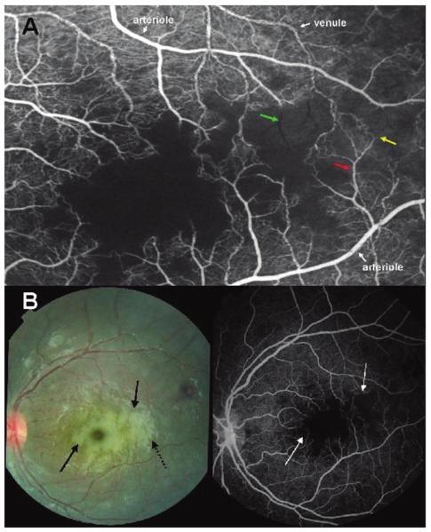
Figure 2.
A, Fluorescein angiogram image of the macula of a child with cerebral malaria showing large areas of capillary nonperfusion (center of picture). Arteriolar and venous circulation have been labelled to illustrate mottling of the blood column, which is most dense in the venules, but also occurs in the arterioles (red arrow). Vessel occlusion is widespread on the border of areas of nonperfusion (resulting in “pruning,” yellow arrow). “Ghost vessels'”can be seen (green arrow); these are nonperfused vessels, the contents of which are blocking the background choroidal fluorescence. B, Paired color retinal photograph and fluorescein angiogram of the same macula. The paired images show the topographical matching between areas of retinal whitening (solid black arrows) and areas of capillary nonperfusion (white arrows). The dark disc at the center of the largest area of macular whitening is the unaffected foveola. Dashed black arrow, artefactual glare overlying retinal whitening.

