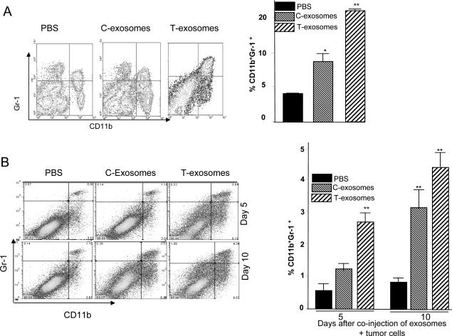
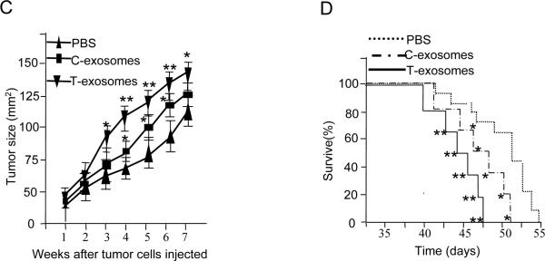
Figure 1. CD11b+Gr-1+ cells induced by T-exosomes promote tumor growth and progression.
7-week old female BALB/c mice (n = 10) were injected intravenously twice weekly for three weeks with exosomes isolated from TS/A tumor removed at day 21 post tumor cell injection (T-exosomes) or exosomes isolated from the supernatants of 36 h cultured TS/A tumor cells (C-exosomes) (100 μg/mouse) . One day after the final injection, the expression of CD11b+Gr-1+ markers on spleen cells was determined using FACS (A, left panel) and the percent of spleen CD11b+Gr-1+ cells was calculated as described in the Material and Methods. Data represent the mean ± SEM of five replicates (A, right panel), * p < 0.05, **p < 0.01. 7-week old female BALB/c mice (n = 5/group) were subcutaneously co-injected with TS/A cells (5×10⁵) and T-exosomes (50 μg). At day 5 and 10 post injection tumors were removed. Collagnase digested single cell suspensions of tumor tissue were stained with anti-CD11b and anti-Gr-1 antibodies, and the percent CD11b+Gr-1+ cells was determined using FACS (B left panel). Data represent the mean ± SEM of five replicates (B, right panel), **p < 0.01. CD11b+Gr-1+ cells were FACS sorted from spleen of naïve BALB/c mice treated with PBS or from naïve BALB/c mice treated with T- and C- exosomes as described in figure legend 1A. The sorted cells were then mixed with TS/A tumor cells (CD11b+Gr-1+ cells : tumor cells = 1:3). Seven-week old female BALB/c mice were injected subcutaneously in the mammary fat pads with TS/A tumor cells alone (1.2×10⁵) or premixed with spleen CD11b+Gr-1+ cells. The growth of the implanted tumor was measured throughout a 50-day period (C). Each point represents the mean tumor size ± SD of 5 mice from each group. The mortality of mice was calculated through day 55 (D). *p < 0.05; **p < 0.01.


