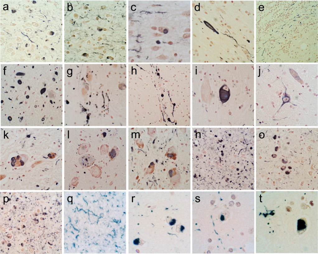
Figure 1.
Photomicrographs depicting immunohistochemical staining for α-synuclein in the brain regions investigated. Positive immunostaining is black; the counterstain is Neutral Red. (a–e) The olfactory bulb and tract. The anterior olfactory nucleus of the olfactory bulb is shown in a–c; both neuronal perikaryal inclusions as well as fibers are present. An enlarged, abnormal neurite within the olfactory tract is shown in d. Positive fibers coursing in parallel array through the internal plexiform layer are shown in e. (f–j) The anterior medulla. The dorsal motor nucleus of the vagus nerve is shown in f, the raphe nucleus in g and i, the internal tract of the IXth nerve in h and the lateral reticular nucleus in j. (k–m) The locus ceruleus in the pons. (n–p) The amgydala. (q) The cingulate gyrus. (r) The middle temporal gyrus. (s) The middle frontal gyrus. (t) The inferior parietal lobule.

