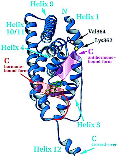
Figure 5.
Inferred coactivator-binding surface. View of hormone-bound hERαLBD and residues important for transactivation as determined by mutagenesis. The active (red) and inactive (magenta) positions of helix 12, red and magenta, are based on the published figures of the hormone- and antihormone-bound hERαLBD structures (1). The domain is oriented as in Fig. 1B. Drawn with ribbons (29).

