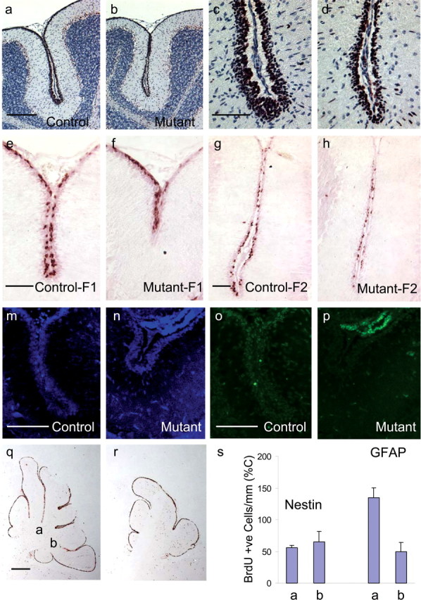
Figure 8.

Granule cell precursor proliferation is impaired in the ilkloxP/loxP; gfap-Cre and ilkloxP/loxP; nestin-Cre mouse brain. a–d, Immunodetection of PCNA is shown in sagittal cerebellar sections of P14 mice. Anti-PCNA antibody was detected by a peroxidase-labeled secondary antibody in combination with Nova Red substrate (reddish brown). Sections have been counterstained with hematoxylin (blue). Proliferative defects occur in the EGL of the folia of mutant mice (b) compared with littermate controls (a). A higher-magnification view of these same folia are represented in c and d. e–h, Representative BrdU labeling at P14 from two different folia labeled F1 and F2 in mutant mice (f, h) and their littermate controls (e, g) are provided. BrdU labeling revealed strong proliferative defects within the cerebellar folia of mutant mice (f, h) compared with littermate controls (e, g). m–p, TUNEL of the EGL of gfap-Cre mice at P14 (o, p) and corresponding areas stained for DAPI (m, n). TUNEL of ilkloxP/loxP; gfap-Cre mice at P14 (p) did not appear different from control (o). q, r, Sagittal sections of nestin-Cre mutant mice (r) and littermate controls (q) at 10 d postnatal were pulsed with BrdU 2 h before the animals were killed and later stained for BrdU (reddish brown). A significant reduction in the number of proliferating cells in the EGL was observed, partly attributable to the loss of folia depth (r). s, gfap-Cre and nestin-Cre mice were injected with BrdU at 10 d postnatal and were killed 2 h later. The number of proliferating GCPs were measured in sagittal cerebellar sections by counting the number of cells having incorporated BrdU in the folia indicated (q). Two or more sections were counted per animal (2–3 wild-type and mutant mice). The number of BrdU-labeled cells in ilkloxP/loxP; gfap-Cre mice (GFAP) and ilkloxP/loxP; nestin-Cre mice (Nestin) along the cerebellar surface adjacent to the primary and secondary fissures are quantified per millimeter of cerebellar surface. This value is expressed as a percentage of the number of BrdU-labeled cells per millimeter cerebellar surface in the littermate control animals. Scale bars: a, 200 μm; c, e, g, m, o, 50 μm; q, 500 μm.
