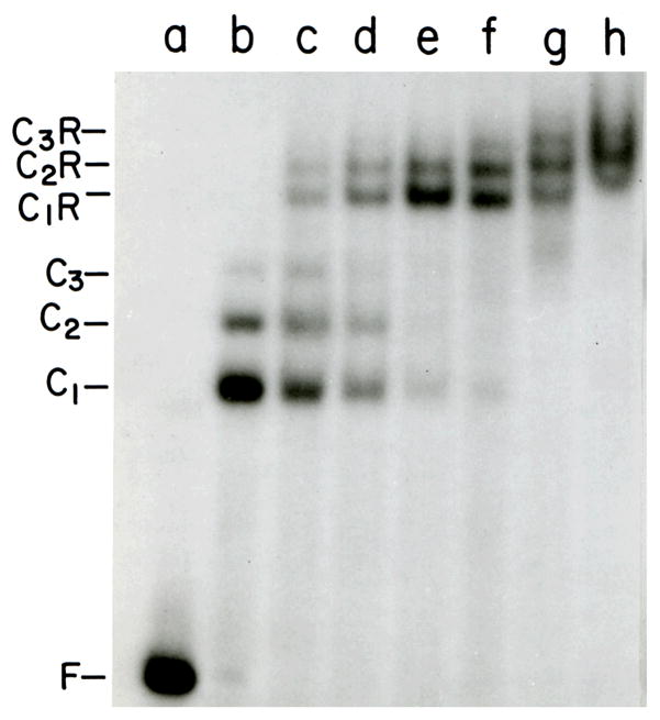
Figure 4. Titration of 1:1, 2:1 and 3:1 CAP-lac promoter complexes with lac repressor.
All samples contained a 214 bp E. coli lac promoter-operator DNA90 (3.7 × 10⁻¹⁰M). Samples b-h contained CAP protein (7.1 × 10⁻⁹M). Samples c-h contained lac repressor at 0.7, 1.5, 2.2, 2.9, 3.6 and 7.3 × 10⁻⁹M, respectively. The binding buffer was 10 mM Tris (pH 8.0 at 20°C), 1 mM EDTA, 50 mM KCl, 20 μM cAMP. Electrophoresis was carried out at room temperature in a 5% w/v polyacrylamide gel run in 45 mM Tris-borate (pH 8.0), 2 mM EDTA, 20 μM cAMP. Symbols: F, free DNA; C1, C2 and C3, complexes with 1, 2 and 3 CAP dimers bound per DNA molecule, respectively; C1R, C2R and C3R, complexes with one repressor tetramer and 1, 2 and 3 CAP dimers bound per DNA molecule, respectively.

