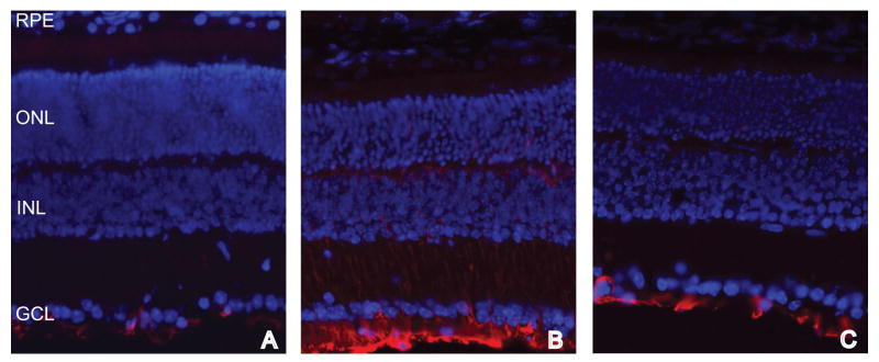
Figure 6.
Fluorescence micrographs of normal (A), untreated rds (B), and EPO treated rds (C) retinas labeled with anti-GFAP (red) and DAPI (blue). GFAP labeling is present only in the Müller endfeet in the normal and EPO-treated rds retinas. In contrast, GFAP labeling is present in the Müller cell endfeet and in processes extending through the retina in the untreated rds retina.

