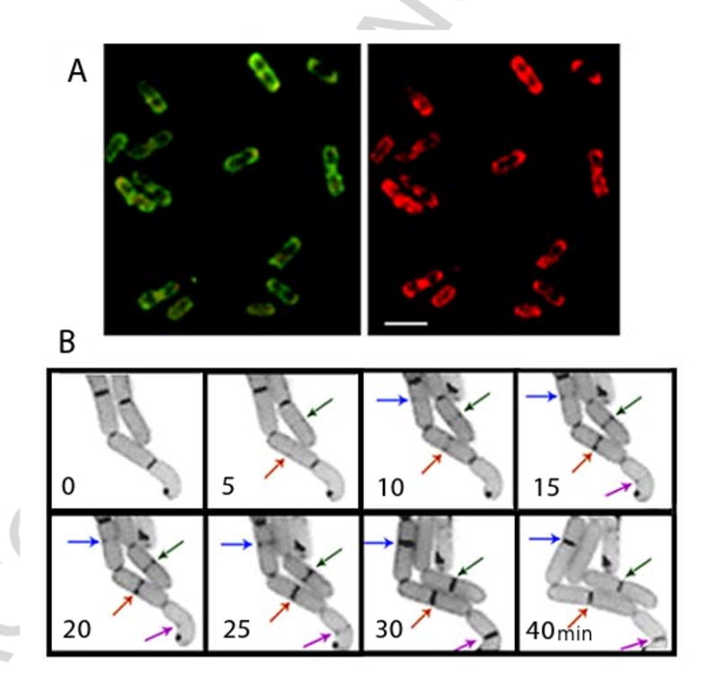
Fig. 1.

Staining of living wild type E. coli cells with fluorescent dye NAO revealed dynamic localization of CL enriched membrane domains. (A) Deconvoluted images of an optical section of cells grown in LB media supplemented with 200 nM NAO. Excitation was at 490 nm and emission was at either 528 (left) or 617 (right) nm; bar, 3 μm. (B) E. coli cells stained with NAO growing on a thin layer of LB agar on a microscopy glass slide. Images were taken with time intervals as indicated. Arrows point to CL domains as they appeared in the mid-cell areas of individual cells and further evolved into complete septa structures enriched in CL. (Fluorescent microscopy was performed by Lu Yang and Eugenia Mileykovskaya)
