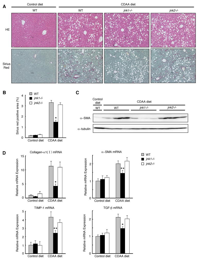
Figure 1. jnk1−/− mice are resistant to CDAA diet-induced liver fibrosis.
Wild-type, jnk1−/−, or jnk2−/− mice were fed a CDAA diet (n = 6–8) or a control diet (n = 4–6) for 20 weeks. (A) HE staining of liver sections is shown (upper panel). Collagen deposition was evaluated by Sirius red staining (lower panel). (B) Sirius red positive area was quantified. (C) Protein expression of α-SMA and α-tubulin as loading control in whole liver extracts was analyzed by Western blot analysis. (D) Hepatic expression of collagen-α1(I), α-SMA, TIMP-1, and TGF-β mRNA was measured by quantitative real-time PCR and normalized to 18S mRNA expression. Values are mean±standard error. *P<0.01, **P<0.05 (jnk1−/− vs wild-type). WT, wild-type.

