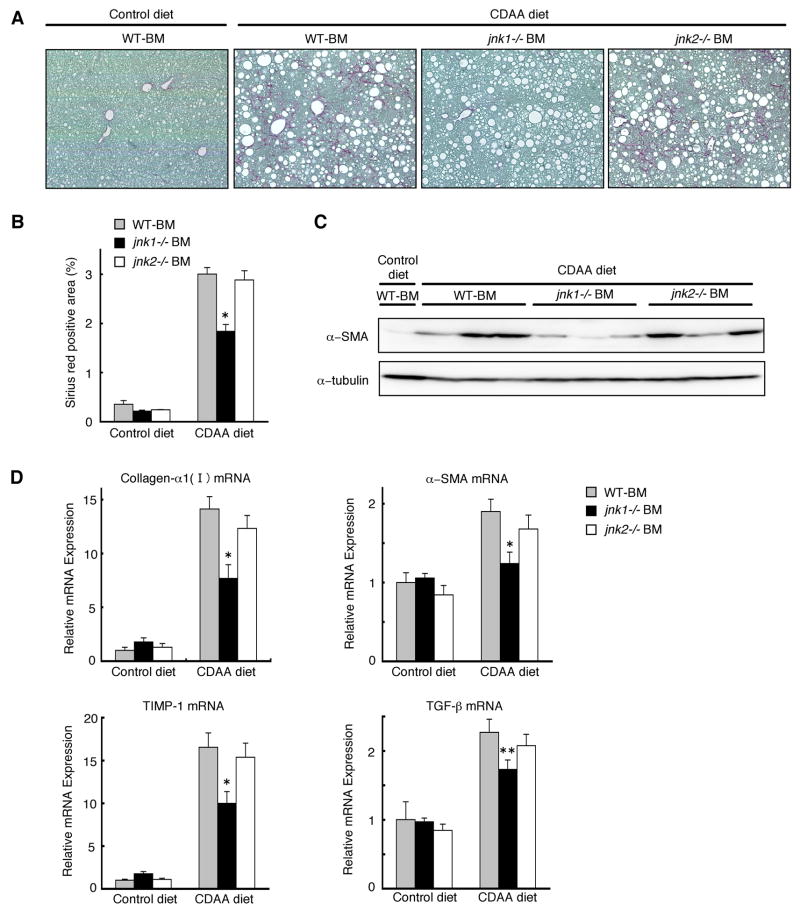
Figure 7. jnk1 deletion in hematopoietic cells decreases CDAA diet-Induced liver fibrosis.
Chimeric mice with wild-type, jnk1−/−, or jnk2−/− bone marrow cells were fed a CDAA diet (n = 8–10) or a control diet (n = 4–6) for 20 weeks. (A) Collagen deposition was evaluated by Sirius red staining. (B) Sirius red positive area was quantified. (C) α-SMA and α-tubulin expression in whole liver extracts were analyzed by Western blot analysis with antibodies against α-SMA and α-tubulin. (D) Hepatic expression of collagen-α1(I), α-SMA, TIMP-1, and TGF-β mRNA was measured by quantitative real-time PCR. Values are mean±standard error. *P<0.01, **P<0.05 (jnk1−/−BM vs WT-BM). WT-BM, chimeric mice with wild-type bone marrow cells; jnk1−/− BM, chimeric mice with jnk1−/− bone marrow cells; jnk2−/− BM, chimeric mice with jnk2−/− bone marrow cells.

