
Figure 2. Ethanol withdrawal-induced neurodegeneration is tPA- and plasminogen-dependent.
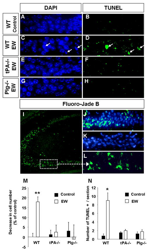
Wild-type, tPA -/- or plasminogen -/- mice were sacrificed two days following EW. EtOH-naïve mice of the same genotype served as controls. Cell loss was determined by counting cell number in the CA1 region of the hippocampus using DAPI-stained sections. To confirm cell death the above sections were processed for detection of broken DNA strands with TdT-mediated dUTP nick-end labeling (TUNEL) method. Analysis of the CA1 region revealed that the number of cells decreased in the wild-type mice (A, C, M) but not in tPA -/- (E, M) or plasminogen -/- animals (G, M) two days after EW. Similarly, we did not observe TUNEL-positive cells in CA1 region of tPA- or plasminogen-deficient mice (F, H, N) typically seen in wild-type mice after EW (B, D, N; arrows in D). Staining using another marker of neurodegeneration, Fluoro-Jade B, confirmed the presence of bright green damaged cells (as seen in the kainic acid-injected positive control; I, L) in the CA1 region in wild-type (J) but not tPA-/- mice (K) two days after EW. Altogether these results demonstrate that EW-mediated neurodegeneration is tPA- and plasminogen-dependent. Sections derived from EtOH-naïve tPA-/- or plasminogen -/- mice did not show any signs of neurodegeneration and are not shown, but are included in quantification in M and N. * p<0.05; ** p<0.01; n=4-5 per group. The results are presented as mean ± SEM.