
Figure 3. Glutamatergic neurons are predominant cell type affected by ethanol withdrawal in the hippocampus.
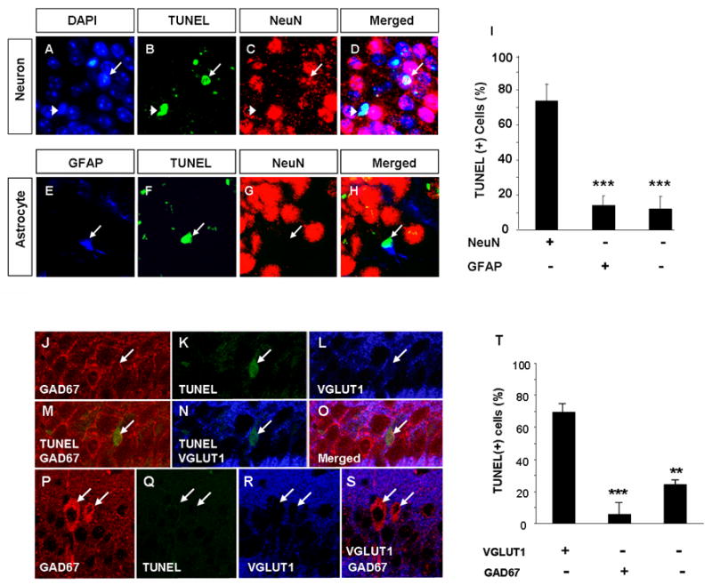
To investigate which cell type is primarily affected by EW we analyzed the phenotype of TUNEL-positive cells in the CA1 region of wild-type mice two days after withdrawal. To this end we performed co-labeling using antibodies against neuron-specific antigen NeuN and astrocyte-specific antigen glial fibrillary acidic protein (GFAP). Panels A-D show two TUNEL-positive cells with chromatin condensation visualized by the DAPI staining. One of these cells (arrowhead) colocalized with NeuN and therefore was identified as a neuron. The second cell (arrow) was NeuN-negative. Panels E-H show a GFAP-positive, damaged astrocyte (arrow). We found that 74.4% of dying cells were neurons (NeuN-positive) and 14.6% were astrocytes (GFAP-positive; quantified in I). Immunohistochemistry for VGLUT1 (a marker of excitatory neurons) and glutamic acid decarboxylase-67 (GAD67; a marker for inhibitory interneurons) in conjunction with TUNEL staining revealed that the majority of dying cells were VGLUT1-positive and GAD67-negative excitatory neurons (J-O). GAD67-positive cells (bright red in P as opposed to punctuate red GAD67-positive synaptic buttons found on most CA1 pyramidal neurons; see J) were TUNEL-negative (P-S). Quantification of the above results shown in T. *** p<0.001. The above estimation was performed using paraffin sections containing CA1 regions of four mice. The results are presented as mean ± SEM.