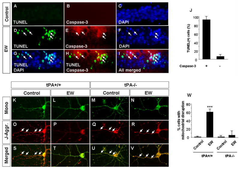
Figure 4. Ethanol withdrawal-induced neuronal death is caspase-dependent and accompanied by mitochondrial dysfunction.

Upper panels Wild-type mice received either ethanol free diet (A-C) or ethanol containing diet followed by EW (D-I). Ethanol withdrawal resulted in neurodegeneration in the CA1 region of the hippocampus as indicated by the presence of TUNEL-positive cells (D, G, H, I). Double immunohistochemistry revealed that the majority of dying cells spatially and temporarily coincided with activation of caspase-3 (arrows in E, H, I and quantified in J). Lower panels. Wild-type (K, L, O, P, S, T) and tPA-/- (M, N, Q, R, U, V) hippocampal neurons were subjected to ethanol withdrawal and mitochondrial integrity was investigated by J-aggregate staining. EW resulted in mitochondrial disruption in wild-type neurons as evidenced by disappearance of red J-aggregates (P, T). In contrast, tPA-/- neurons were protected against EW-induced injury (arrows in R, V; quantified in W). *** p<0.001. The above experiments were repeated at least four times with similar results. The results are presented as mean ± SEM.
