
Figure 5. tPA-/- neurons in culture are resistant to EW-induced neurodegeneration.
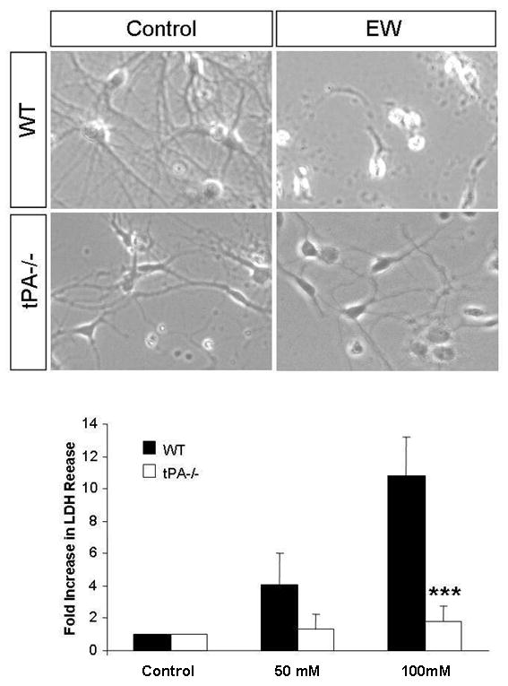
To determine whether ethanol mediates its' effects at the cellular or rather circuit level, we investigated the effects of EW on the survival of dissociated wild-type and tPA-/- hippocampal neurons in culture. Neurons were exposed to 50 or 100 mM ethanol for 3 days and subject to withdrawal. Bright field images of neurons exposed to 100mM ethanol revealed signs of cell death (rounded cell bodies, short neurites) in wild-type neurons undergoing EW, where as tPA-/- neurons were resistant to EW-induced neurodegeneration (upper panels). EW-induced neurodegeneration was quantified using the LDH assay (lower panel). Withdrawal from 50, and 100 mM ethanol resulted in a dose-dependent increase in LDH release from wild-type neurons (n=4) whereas tPA-/- neurons did not show a significant increase in LDH release (n=5). ***p<0.001. The results are presented as mean ± SEM.