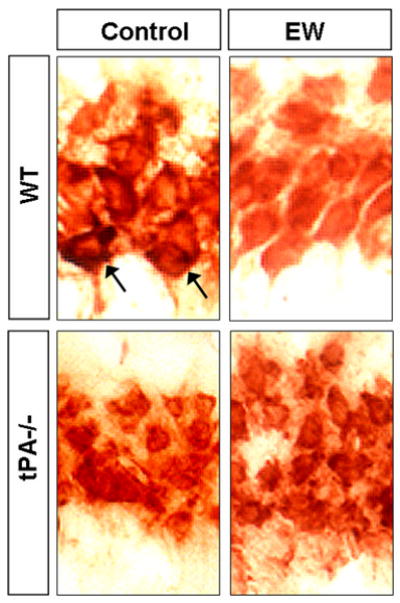
Figure 6. EW-induced neurodegeneration correlates with tPA-dependent degradation of laminin.

To investigate if EW-induced neurodegeneration correlates with laminin degradation we assessed laminin levels by immunohistochemistry in the hippocampal CA1 region of wild-type and tPA -/- mice during EW. EtOH-naïve mice of the same genotype served as controls. Laminin was localized mainly around cell bodies. In wild-type mice a marked decrease in laminin levels was observed six hours after EW in the CA1 (strong pericellular staining indicated by arrows), the region that degenerates two days later. Laminin degradation was not observed in tPA-/- mice, which were also resistant to neurodegeneration. These results suggest that degradation of laminin is tPA-dependent and could be responsible for EW-induced neuronal death. This experiment was repeated four times with similar results.
