Figure 1.
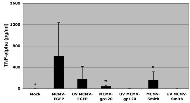
The efficient activation of DC by MCMV depends on the expression of immediate early and early genes as well as transgenes. A: IL-12, IL-10, IL-6, and TNF-α induction by MCMV-EGFP-infected and LPS (1μg/ml) treated DC at 24 and 48 h after infection. Representative results from one donor are shown. The experiment was repeated at least three times with cells derived from different donors and similar results were obtained. B: TNF-α induction by MCMV-EGFP, MCMV-gp120, MCMV-Smith and UV-inactivated MCMV-infected DC at 12 h after infection. Each column represents the data of four individual experiments from four donors (means + SD). An asterisk indicates a statistically significant difference (p-value < 0.05; ANOVA) compared to MCMV-EGFP group.
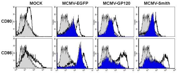
C: Up-regulated expression of CD80 and CD86 in MCMV infected DC at 48 h after infection. Gray filled histogram: PE labeled IgG1 isotype control antibody; Open histogram: Specific staining with either PE labeled CD80 or CD86 antibodies for live virus-infected cells. Blue filled histogram: Specific staining with either PE labeled CD80 or CD86 antibodies for UV-inactivated virus-infected cells. Dead cells were excluded from the flow cytometric analysis by using propidium iodide staining. The experiment was repeated twice using Mo-DC derived from two separate donors and similar results were obtained.


